Keratoconus Treatment
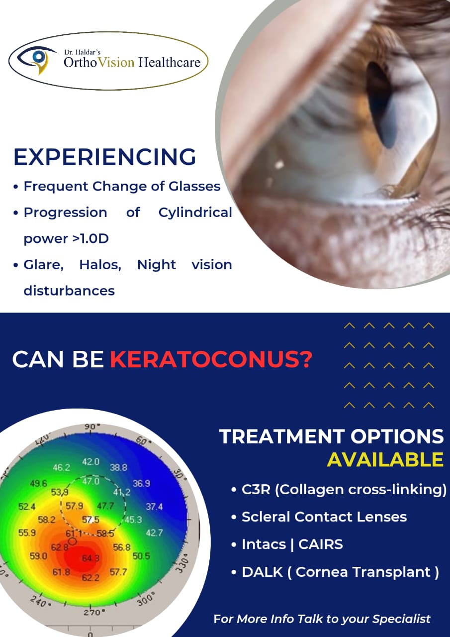
Keratoconus is an ectatic corneal degenerative disorder in which the cornea begins to weaken and starts thinning and steepening(bending) causing visual disturbances. As the name suggest the spherical cornea becomes conical in shape, hence ‘keratoconus’-(kerato-cornea, conuc-conical) The clear transparent cornea focuses light beams onto the retina. Any anomaly in its shape causes visual disturbances.
How do eye doctors diagnose Keratoconus?
Your ophthalmologist might utilize various instruments to make a finding of keratoconus, including:
- Still Lamp Evaluation
- Keratometry
- Corneal Topography ( Pentacam / Orbscan )
How do eye doctors treat Keratoconus?
In the early stages of keratoconus, the main treatment given by the Eye specialists might require the use of glasses to address your vision. In later stages vision may not be very clear with glasses.
That’s when you might need special contact lenses called Rigid/ Hard contact lens. These are available in various types depending upon stage and steepening of cornea.
- RGP(rigid gas permeable lenses)
- Miniscleral lenses
- Rose k lenses
- Scleral lenses.
Corneal collagen cross-linking(C3R)
- Corneal cross-linking is a procedure that can be done in mild- moderate cases of keratoconus with documented evidence of progression over one year(increase in steepening by 1D)
- This involves treating the cornea with Riboflavin eyedrops for 20-30 minutes (priming procedure) followed by UVA radiation beam exposure for 10-30 minutes to fortify and strengthen the cornea so that it doesn’t bend further.
Intacs/ICRS
- In advanced asymmetric cones(as seen on corneal topography), intacs or intracorneal segments are placed in the cornea to regularise the shape of the cone so that contact lenses would fit better and visual quality would be better. They do not correct the existing refractive error completely but only modify the shape of the cornea.
Corneal transplantation (keratoplasty)
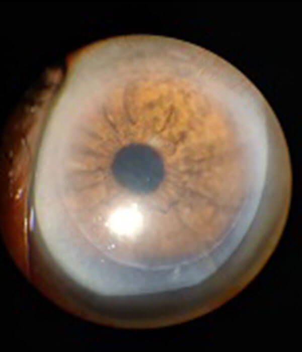
- In advanced keratoconus, such as post hydrops scarring where bowmans membrane layer of cornea is already ruptured due to too much thinning of cornea leaving behind a scar in centre of cornea thus obstructing the visual axis, cornea surgeons may perform a partial thickness or a full thickness corneal graft to replace the scarred cornea with a transparent donor cornea.
- The donor corneal graft is sutured to host corneal rim with 16-20 fine sutures which are subsequently removed after 8-9 months till the donor cornea is fully integrated with host rim. However one can start doing routine activities after a week.
These are the best available options for keratoconus with few modifications in every procedure to maximise visual outcome in our patients.
Which is the best place to get Keratoconus therapy?
If you are suffering from keratoconus(as already diagnosed) or you are frequently changing your glasses or you are not happy with your quality of vision do consult an eye specialist as an increasing cylindrical power may be a subtle sign of early keratoconus. This may be confirmed by clinical evaluation and corneal topography.
For any further queries do contact us at Dr. Haldar’s OrthoVision Healthcare, Noida. We will assist you through expert guidance and communication.
Before & After Gallery

Molten metal burn

After Tenonplasty

Corneal Opacity

After Cornea Transplant

Fuch’s Dystrophy

After DSAEK
Video Archive

Dr. Sridevi Haldar
DNB, FCRS
Cataract,Cornea & Refractive Eye Surgeon
Dr. Sridevi Haldar (Gunda) is an eminent eye surgeon providing expert services to leading city hospitals across Delhi NCR. She has been associated with Dr. Shroff Eye Hospital, Delhi; Venu Eye Institute, Saket and Sharpsight Centre in the past.
Qualification: After graduating in year 2005, she pursued her interest of specializing in the field of Ophthalmology and completed her post-graduation from world renowned, prestigious instate;”